HELLO STEEMIANS!
Today, I'm going to share a little more about me and what I do at my job. A few weeks ago, I had posted this but in spanish, so here is the english version.
I work in a medical center in Caracas, Venezuela; synthesizing radiopharmaceuticals in nuclear medicine for the diagnosis of different diseases, mainly oncological. Below I explain in more detail what it consists of:
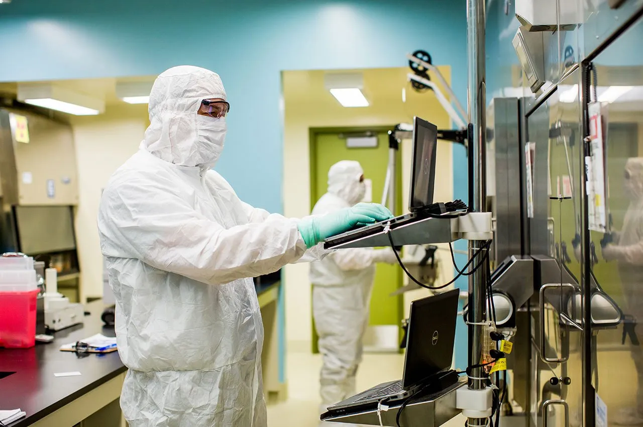
Image source
PET-CT Scan.
Positron emission tomography and computed tomography (PET-CT) is a diagnostic technique used in nuclear medicine. Through it, by intravenous administration of a small amount of a tracer radiopharmaceutical, metabolic images of the interior of the human organism can be obtained.
This technique detects the metabolic activity of the cells you wish to study/evaluate. For example, you can determine the sugar consumption of certain cells. For this reason, PET-CT is used mainly for the diagnosis and monitoring of patients with cancer, since the tumor cells have a different metabolism than healthy cells.
The radiopharmaceutical circulates through the body and adheres to the tumor cells, where it begins to emit a small amount of radiation. This radiation is detected by the PET machine, which produces an image in those areas of the body where the radiopharmaceutical has accumulated, which may correspond to tumors stand out.
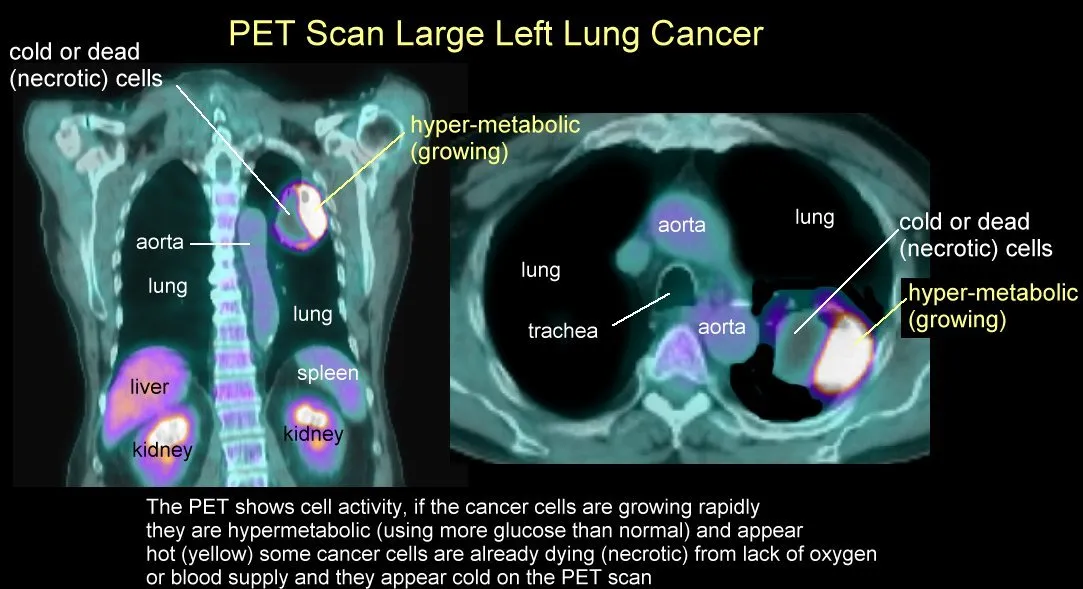
Image source
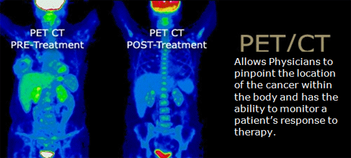
Image source
Preparation of tradiopharmaceutical tracer.
There are several radiopharmaceuticals used in nuclear medicine, which are used according to the technique and the medical area required by the study (cardiology, neurology, oncology, among others). They are chemical compounds whose radioactive half-life is relatively short but enough to carry out the specific study. In our medical center, we used 18-FDG (18-fluorodeoxyglucose) as radiopharmaceutical tracer for PET-CT scan.

Image source
The 18-FDG molecule differs from the glucose (sugar) molecule only in the fluorine 18 atom that does not have the latter, so the human body "sees" it and metabolizes it as if it were sugar. On the other hand, fluorine 18 (18F) is a radioactive isotope of fluorine atom, that is, it emits energy in the form of ionizing radiation, which is what the tomograph captures.
Two steps are necessary to prepare the 18-FDG ; first of them involves a nuclear reaction to obtain 18F and the second step is a chemical reaction that inserts the isotope 18F into the glucose precursor molecule (ultra pure mannose triflate).
The 18F isotope must be created (nuclear reaction) first as fluoride (18F-) in a big machine called cyclotron, wich is a particle accelerator. This is carried out by proton bombing (H +) in water enriched with 18O, which results in a synthesis reaction of radiolabeled (18F-) in the form of hydrofluoric acid, HF, (step that can take up to 2 hours), by the following nuclear reaction: 18O (p, n) 18F.
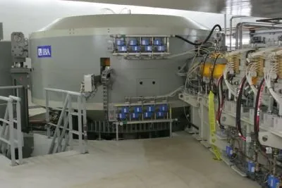
Image source
The following video shows, in a very didactic way, how a cyclotron works . Courtesy of Depinxi Youtube channel.
After 18F is obtained in its fluoride form, it must be inserted into the glucose precursor molecule, mannose triflate; as discussed above in order to obtain 18-FDG, through a chemical reaction called bimolecular nucleophilic substitution (SN2) . This reaction takes 30 additional minutes and is carried out by another equipment that performs it in an automated way. This maquine is called synthesis module. Chemical reaction is schematized in the following figure:
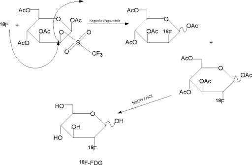
Image source
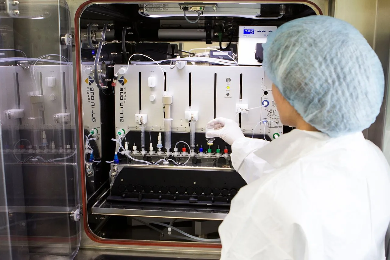
Image source
Once the 18-FDG radiopharmaceutical is obtained, it must be submitted to strict quality controls to be suitable for the administration of patients. To do this, a small amount of 18-FDG sample is taken, and tests are carried out in order to determine pH, the concentration of remanent salts, side products and byproducts from the chemical reaction and excess reagents and solvents. Moreover, is necessary to know the radiochemical purity of the drug, its half-life and finally to carry out microbiological tests. All this tests must be done promptly and before the injection of patiens, because the radiotracer drug obtained, 18FDG, has a short half-life which is 110 minutes.
If the radiopharmaceutical does not pass one of the quality controls, it is not suitable for anyone's injection. Several aboratory techniques are neccesary, such as gas chromatography, thin layer chromatography, pH measurementt, and tests for the control of endotoxins; also gamma radiation detectors and dose calibrators are used.
Image source
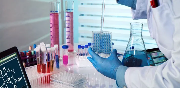
Once the quality controls have been approved, the radiopharmaceutical must be distributed either for external delivery or to be administered in the nuclear medicine service of our medical center. Depending on the weight of the patients and the time they would be injected, small fractions of the drug will be extracted from the container that contains the entire volume of the radipharmaceutical solution and placed in syringes.
Fractionation of the radiopharmaceutical drug is done manually, under a sterile laboratory hood and following the daily safety rules to avoid overradiation. At all times, the solution container, as well as the transport syringes of the drug will be under lead armored containers, since lead as material, together with a good thickness of it, protect the operator from exposures to radioactive energy.
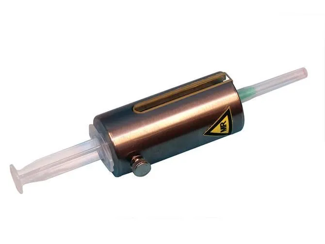
Image source
Prior to the injection of the radiotracer, patients must comply with some medical indications. Namely, they must be at least 6 hours fasting, have the capillary glycemia below 140 mg/dL and comply with other specific treatments depending on the reason for the study and their pathology. This in order for the study to go unchanged.
After the injection, patients must remain at rest in a dark room, very calm and relaxed for approximately 45 minutes, in order to allow time for their body to metabolize the drug throughout its anatomy. After this time, the patients are ready to go to the PET-CT equipment, where they will remain lying in the position indicated by PET operator for 25 to 50 minutes:
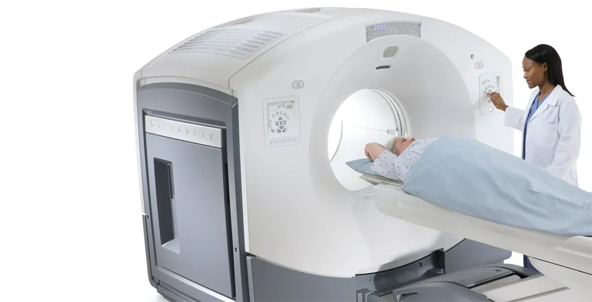
Image source
Finally, the radiologist processes the images in a computer program and the imaging doctor is responsible for evaluating them and making a final report, which is delivered to the patients along with the images obtained so that their main doctors can give them the diagnosis and treatment to follow.
More about me:
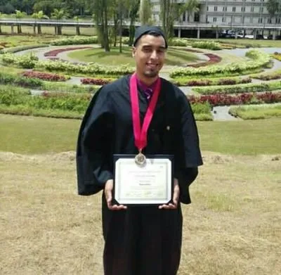
I graduated with a degree in Chemistry from the Central University of Venezuela in 2009, which is why I was hired to work in this area. I am also one of the only three people in Venezuela who do this work, since only at the medical center where I'm working , the 18-FDG is prepared in this country.
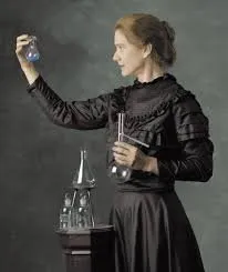
This post is dedicated to Marie Curie (maybe @curie? hahaha), Nobel Prize in Physics in 1903, pioneer in the field of radioactivity, discovered the radio and polonium elements:
Original text by myself (@oscardice), supported in reference 1 and in my work experience.
For more information about PET-CT Scan, you can watch the following video, courtesy of Queensland X-Ray Youtube channel:
Reference:
- López-Durán, Fred Alonso; Zamora-Romo, Efraín; Alonso-Morales, José Luis; Mendoza-Vásques, Guillermo. "TOMOGRAFÍA POR EMISIÓN DE POSITRONES: LOS NUEVOS PARADIGMAS", Tip Revista Especializada en Ciencias Químico-Biológicas, vol. 10, núm. 1, junio, 2007, pp. 26-35. Universidad Nacional Autónoma de México Distrito Federal, México.
Due to privacy and security policies of the company where I am employed, I do not have permission to publish photograph and information of my workplace, so all the photos placed here are referential and as close to reality.

