Hello? I am @radiologist.
Do you know that the heart gets a CT scan?
Recently, a lot of cardiac CT have been performed for health screenings or the evaluation before and after the operation. Today, I will talk about cardiac CT.
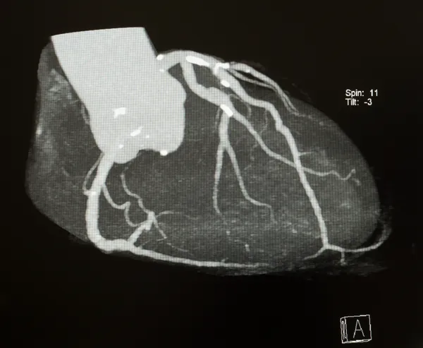
A 3D reconstruction image of cardiac CT, the brightest white is a calcified plaque of blood vessels.
The principle
Your heart is working hard at this moment. In a nutshell, the heart is pumping blood into the body and the lungs. If there is movement, the quality of the CT image will be degraded, like taking a picture of moving stuff.
Then, is it okay to get a heart (moving and pumping now!) CT scan? Although CT scan time has been shortened recently, it is still too hard to visualize the heart in one second (the time needed to get general CT scans).
So, how do you get an image of your heart?
The solution is simple. As the heart keeps pulsing at a regular cycle, by capturing only a specific point in the cycle and taking a small bit of image over several cycles, a complete heart can be seen. It is like that you put a lot of pieces of puzzle to get a single picture.
Visualizing moment should be when the movement of the heart is the smallest. For taking a image at this moment, the electrocardiogram(ECG) is used. This technique is called ECG gated CT. Then, when is the moment?
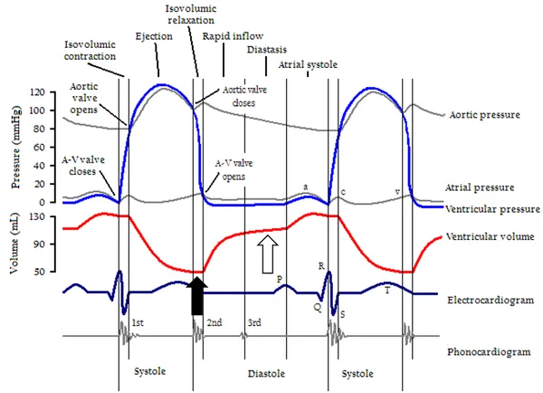
This picture shows the cycle of the heart. Depending on the heart rate, the periods of low motion are slightly different. At a slow heart rate of 70 or less, the image is acquired in ‘diastasis’ period (white arrow), and at a rapid heart rate of 70 or more, the image is acquired in ‘isovolumetric relaxation’ period (black arrows) at the end of the systolic phase.
How do you catch the best moments in CT? There are two main ways. All are caught through the patient's ECG (you can get ECG in the CT machine). Two methods are prospective(forward) synchronization and retrospective(backward) synchronization.
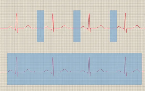
The blue box is the moment of the CT scan, The above is prospective(forward) synchronization, and the bottom is retrospective(backward) synchronization.
Prospective(forward) synchronization is a method of obtaining the image after a delayed time when the R (highest peak) wave arrives in ECG. In other words, it is a way to get a scan at the exact timing through ‘predicting’ in advance. This method gets a CT scan only when it is absolutely necessary, so radiation dose is low. However, if there is arrhythmia (irregular heartbeat), it is difficult to predict the exact time, so the quality of the image may be degraded.
In retrospective(backward) synchronization, all the images are taken regardless of the cardiac cycle. After that, you can reconstruct only the images at specific points by matching them with the ECG. Of course, because the images of entire cycle are taken, there is a lot of radiation exposure and the CT device may be overloaded. However, as you get images of various cycles, you can only pick out the best images. Moreover, you can evaluate the movement of the heart muscle, because there are the images of continuous movement. In addition, it is less affected by arrhythmia.
Regardless of the methods, it is based on the ECG to acquire and reconstruct the image at the best time, finally waiting for the radiologist to read it.
Pretreatment
A slow heart rate can result in less movement of the coronary arteries and a better image. So if the heart rate is fast, a heart rate can be artificially reduced. If your heart rate exceeds 65 to 70, you can use a beta-blocker to control your heart rate. In addition, nitroglycerin(NTG) can be used because coronary arteries must dilate to be well evaluated by CT. NTG should not be used if you have low blood pressure, cardiac abnormality, glaucoma, or if you are taking Viagra(it’s also vasodilator like NTG).
The Use
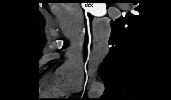
A Heart CT Scan - Multi-planar reconstruction of the right coronary artery (Multiplanar reconstruction, MPR : You can understand that you look at the blood vessels in a straight line though reconstruction program.
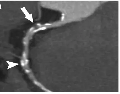
A Heart CT Scan - Right coronary artery MPR images show calcified plaque and non-calcified plaque (look at the arrow).
The most basic use is assessment of coronary arteries(supplying blood to the heart). In the past, coronary angiography (inserting catheter into blood vessels and evaluating through a contrast media) was the only method, but now cardiac CT is somewhat replaceable. It is often used as medical check-up or screening test (chest pain is the main cause) and can be taken when an acute myocardial infarction (coronary artery obstruction) is suspected.
However, cardiac CT is not superior to angiography. So if the coronary artery stenosis seems to be severe on CT, coronary angiography can be used to evaluate accurately and to treat coronary artery stenosis(angioplasty or stent insertion). Often, stenosis on cardiac CT looks severe, but it does not look too severe on coronary angiography. In other words, CT tends to overestimate coronary artery stenosis. But cardiac CT has many advantages because it is less burdensome than invasive coronary angiography.
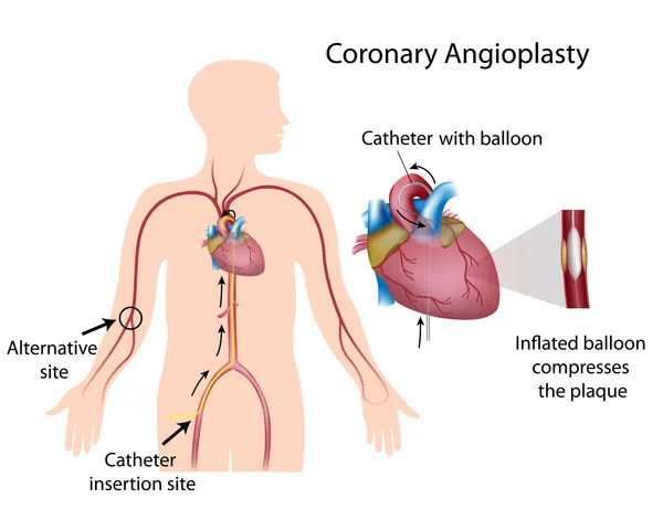
If the heart is abnormal on CT, coronary angiography(CAG) is performed. If necessary, angioplasty or stent insertion can be performed.
In addition, cardiac CT has been largely used to evaluate graft vessels after coronary artery bypass graft (CABG) surgery, the patency of stent, complication after valvular replacement, and great vessels including aorta.
To conclude…
I’d like you to understand cardiac CT a little bit more through this posting. Cardiac CT started late than other body part. However, the performance of CT improves and techniques for reconstruction are evolving rapidly. Therefore, it will be more useful and valuable than before.
Reference
[Korean] 박재형. 심장혈관영상의학. 일조각. 2011.
- Images without source; from Shutterstock.

This work is licensed under the Creative Commons Attribution-Noncommercial-NoDerivatives 4.0 International License.

